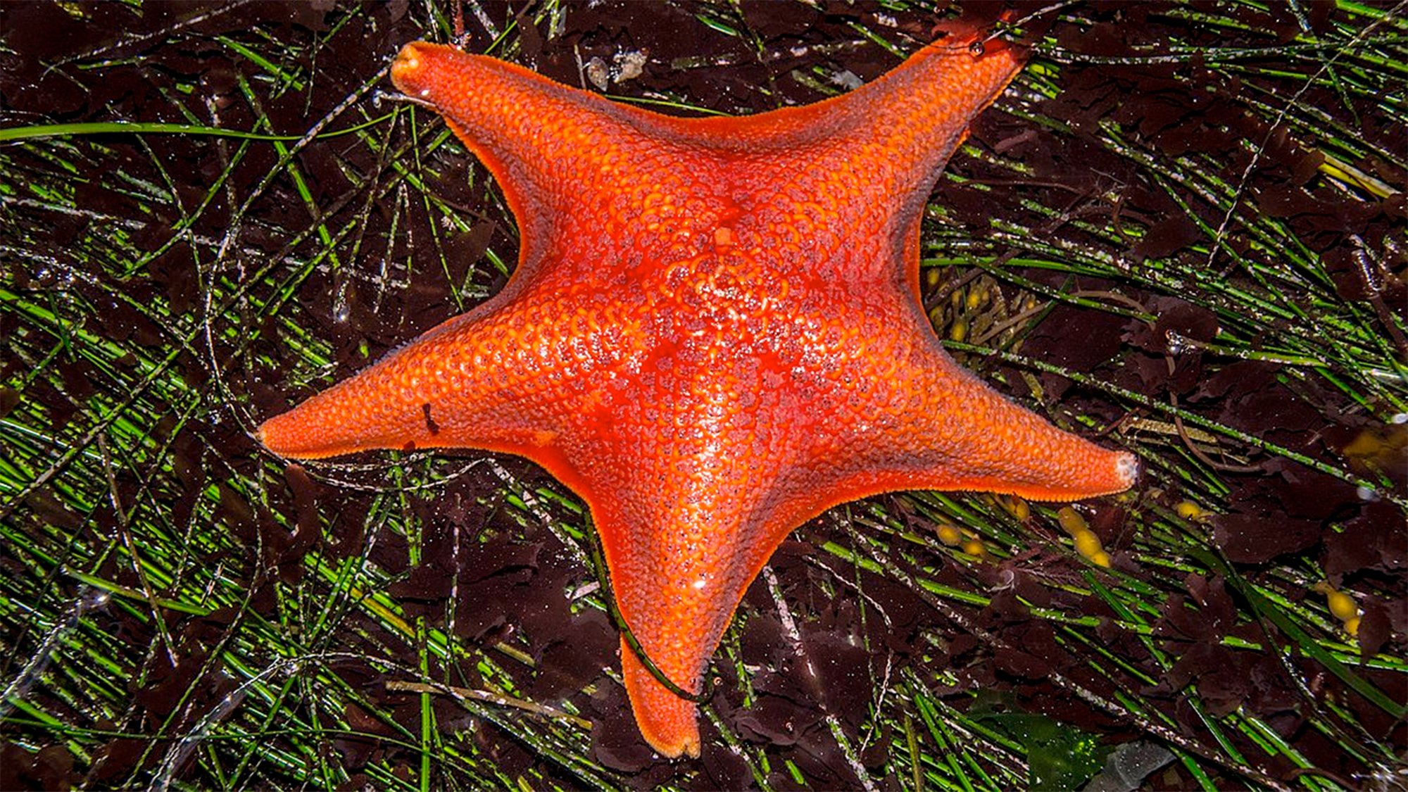

The humble sea star is an ancient marine creature that possibly goes back about 480 million years. They are beloved in touch tanks in aquariums for their celestial shape, spongy skin, and arm suckers. These beautiful five-limbed echinoderms are also helping scientists figure out a crucial life process called tubulogenesis.
[Related: What’s killing sea stars?]
A study published May 9 in the journal Nature Communications, examined this process of hollow tube formation in sea stars that provides a blueprint for how the organs of other creatures develop.
Tubulogenesis is the formation of various kinds of hollow, tube-like structures. in the body. These tubes eventually form blood vessels, digestive tracts, and even complex organs like the heart, kidneys and mammary glands. It is a basic and crucial process that occurs in the embryo stage, and abnormalities during these processes can cause dysfunctional, displaced, or non-symmetrical organs and even regeneration defects in structures like blood vessel.
Little is known about the general mechanisms of the hollow tube formation during embryogenesis since animals all use very different strategies to form these tubular structures.
That’s where the sea star comes in. Their process of tubulogenesis is relatively easy to observe since their embryos are very transparent and can be observed without disturbing them. Not to mention, they breed in large numbers year round. This new study reveals the initiation and early stages of tube formation in the sea star Patiria miniata or bat star.
“Most of our organs are tubular, because they need to transport fluids or gasses or food or blood. And more complex organs like the heart start as a tube and then develop different structures. So, tubulogenesis is a very basic step to form all our organs,” study co-author and cell biologist Margherita Perillo of the University of Chicago-affiliated Marine Biological Laboratory said in a statement.
Not only is the sea star an ideal because of its translucence, the researchers needed an animal that was along the base of the tree of life and evolved before the phylum Chordata– vertebrates including fish, amphibians, reptiles, birds, and mammals, Perillo adds.
Perillo and her colleagues used CRISPR gene editing and other techniques to analyze the gene functions in the sea stars and long time-lapse videos of developing larvae. The team worked out how the sea star generates the tubes that branch out from its gut. From these observations, they could define the basic tools needed for more advanced chordate tubular organs that may have developed. Now, they are getting closer to answering how organisms developed up from one cell into the more complex 3D tubular structures that make up various organisms.
According to Perillo, in some organisms such as flies, “there is a big round of cell proliferation before all the cells start to make very complex migration patterns to elongate, change their shapes, and become a tube.”
[Related: These urchin-eating sea stars might be helping us reduce carbon levels.]
In other animals, including mammals, cell proliferation and migration occur together. The team found that in sea stars, cells can also proliferate and migrate at the same time in order for the tubes to form the way they do in vertebrate formation. The mechanism behind making organs must have already been established at the base or root of chordate evolution, according to the team.
Beyond providing evolutionary insights into organ formation, sea stars can also aid in biomedical research. Perillo found that a gene called Six1/2 is a key regulator of the branching process in tube formation. If Six1/2 is taken out of mice, their kidneys form abnormally, but the mice that lack the gene also resist tumor formation, even if they are injected with tumor cells. Understanding this gene, that is overexpressed in cancer cells, may lead to new ways to study disease progression.
“I can now use this gene to understand not only how our organs develop, but what happens to organs when we have a disease, especially cancer,” said Perillo. “My hope is that, in five to 10 years maximum, we will be able to use this gene to test how organs develop cancer and how cancer becomes metastatic.”